

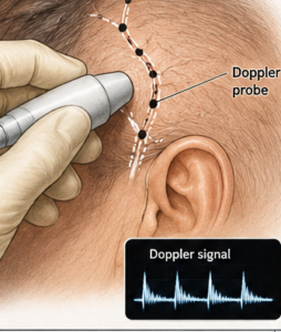
In temporal artery ligation procedures (most commonly for treatment of prominent superficial temporal artery branches, a Doppler ultrasound probe plays an important localization role.
Roles of the Doppler
1. Preoperative Vessel Mapping
- Handheld Doppler identifies the exact course of the superficial temporal artery and its frontal and parietal branches should it not be easily visible at the time of the surgery.
- This is particularly valuable because the artery’s course varies among patients.
- The vessel can be marked on the skin before incision.
2. Localization Through Small Incisions
- Doppler allows the surgeon to pinpoint the artery through which it can be ligated with a very limited incision.
- This minimizes scarring and tissue trauma.
3. Confirmation of the Correct Vessel
- The arterial pulsatile signal confirms that the structure identified is truly the temporal artery rather than a vein or fibrous tissue.
- Particularly useful when determining if there any arterial branches contributing to its prominence that can not be seen.
4. Identification of Multiple Branches
- The superficial temporal artery often bifurcates into frontal and parietal branches.
- Doppler helps determine whether additional branches require ligation to achieve the desired result.
5. Verification of Successful Ligation
- After tying the vessel, the Doppler signal can be reassessed.
- Absence of flow distal to the ligation confirms successful occlusion.
Practical Technique
Most surgeons use an 8–10 MHz handheld Doppler:
- Mark the strongest signal over the artery.
- Make a small incision directly over the vessel.
- Dissect to the artery.
- Place ties proximally and distally.
- Recheck with Doppler to confirm elimination of flow.
For aesthetic temporal artery ligation Doppler-guided localization is the most definitive method that goes beyond seeing and feeling to ensure that flow through the artery has been eliminated.
Case Example

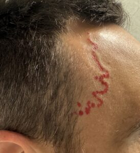 This middle ages male developed a right temporal artery prominence for unknown reasons and desired it ligated. At the time of the procedure, as ir common, the arterial prominence was barely visible. The arterial course was marked along its typical serpiginous course.
 Three ligation points were marked with the possibiolity of a fourth more distal.
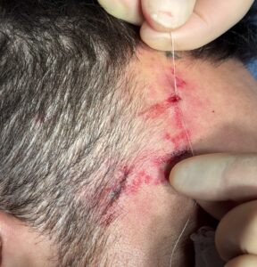
 Under local anesthesia the most proximal site at the edge of the temporal hairline was double ligated.
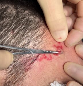 The second or middle ligation location was double ligated after its arterial loop was extracted out of the incision.
 The third location was similarly exposed and ligated.

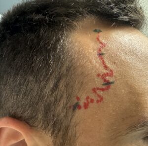
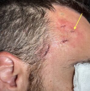 A doppler check was done and a pulsate signal was located distal and more anterior to the second ligation site. It was there ligated as well (yellow arrow).
Discussion
A multi level ligation technique can be very effective for eliminating a prominence of the superficial temporal artery. It has an over 95% success in doing so in hundreds of cases. The key is ensuring by the completion of the procedure that there is no longer any audible arterial signal along the course of the vessel. The use of the Doppler ultrasound has proven invaluable in identifying arterial branches after the typical three ligation points are done that otherwise would have been missed.
The typical number of ligation points needed is usually four. The initial locations are three, proximal, middle at the side of the forehead, and this’ll closer up to where the artery starts to enter the scalp. Once these three ligation points are done a signal in the path of the artery is checked. It is only in rare cases that the three ligation points are satisfactory. Almost always there is a fourth or even a fifth ligation site, identified only through the use of the Doppler, that is necessary for a complete elimination of the signal along the arterial course. It is important in doing the procedure that one takes the time to search for such signals to prevent reoccurrence of any part of the initial vessel prominence.
Case Highlights:
- successful ligation of the prominent, superficial temporal artery, often takes more than three ligation sites.
- The Doppler is most useful in identifying ligation sites after the primary visible ones are completed.
- Do use of the Doppler does not ensure that complete elimination of the prominent temporal artery can be achieved in every case, but it does provide a simple technological tool to achieve better results than can be done with one’s eyes alone.
Dr. Barry Eppley
Plastic Surgeon