
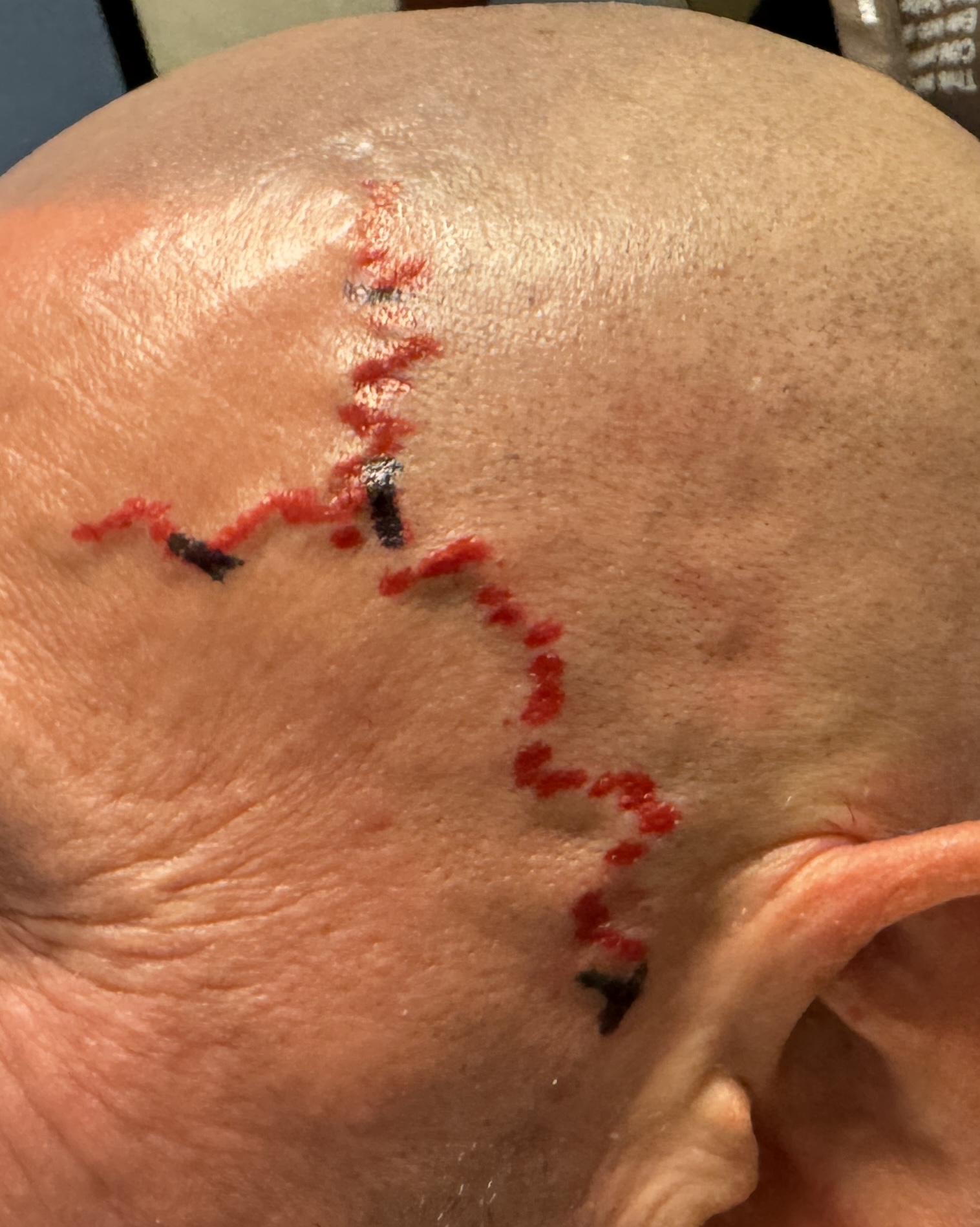

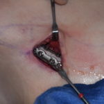
In the treatment of large, prominent superficial temporal artery (STA) in a male patient for cosmetic reduction, the important issue is that simply tying off one visible point will not reliably eliminate the vessel. It requires a multi-level ligation technique for assured effectiveness.
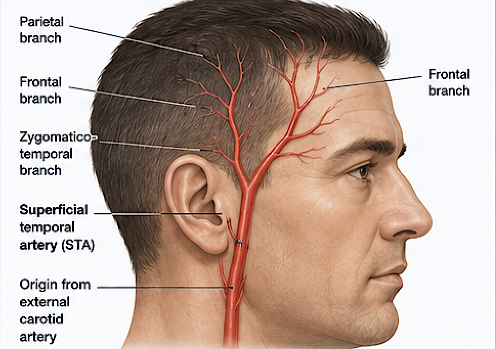 The STA has a collateral-rich network. Its frontal and parietal branches communicate with each other and with supraorbital, supratrochlear, occipital, and contralateral vessels. Consequently, after a single ligation, a prominent segment can potentially continue filling through collateral or retrograde flow. Multilevel ligation is intended to interrupt that filling at more than one point along the abnormal/prominent vascular pathway.
For operative planning, the appropriate levels can be determined just from surface prominence or the visible branching pattern. Intraoperatively, however, a particularly large or unusual temporal vessel should first have its anatomy and flow assessed as the ligations proceed—commonly with an ultrasound doppler —because confirmation of the absence of flow needs to be assured beyond visual or palpable assessments. It can occasionally be surprising how even between two ligation points along an obvious arterial branch how a flow signal may still exist.
Case Example
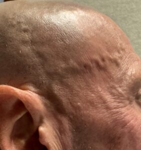
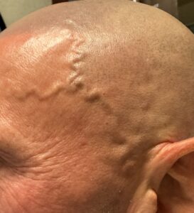 This shaved head male developed very large prominences of the STAs for no known reasons. With his shaved head and their prominences extensive branching patterns were seen with different patterns on each side. The right side had the classically described Y pattern with the split in the mid-temporal region. On the left side the Y split was located more anterior at the edge of the hairline with no branching in the temporal hair bearing area.
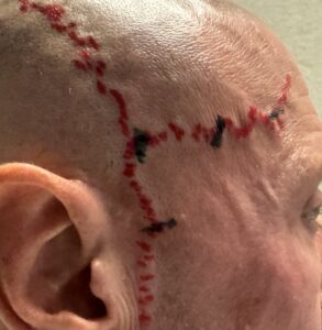
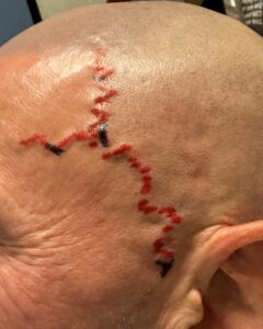 The STA branch patterns and ligation sites were marked.
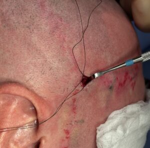

 On both sides a three site ligation technique was done at and beyond the Y configuration.
 Due to the very strong flow and large vessels the main temporal artery was also ligated in front of the ear.
 Confirmation of flow ablation along the arterial prominence was confirmed by doppler assessment.
Discussion
In prominent superficial temporal arteries (STA) visible along the temple/forehead, they can show different branching patterns because arterial anatomy naturally varies from person to person.
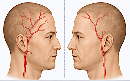 The superficial temporal artery typically travels upward in front of the ear and divides into frontal and parietal branches, but the exact configuration isn’t fixed. Differences can include where the artery divides, how many secondary branches arise, their diameter, and the direction they travel.
A major reason is developmental variation. During embryonic development, blood vessels form through a network that undergoes remodeling. Some channels enlarge and persist while others regress. Small differences in this process produce different adult arterial patterns without necessarily representing anything abnormal.
There are also extensive anastomoses—connections between neighboring arteries—particularly involving branches of the superficial temporal, supraorbital, supratrochlear, and occipital arterial systems. Because these vascular territories overlap, one person’s STA may supply an area through a large obvious branch while another person’s supply may be distributed through several smaller branches.
So you might see patterns such as:
- an early versus late bifurcation of the STA,
- one dominant frontal branch versus several branches,
- prominent secondary or accessory branches,
- different courses across the temple and forehead,
- asymmetry between the left and right sides of the same person.
Prominence of the vessel, regardless of the branching pattern, depends on vessel diameter, skin/subcutaneous-tissue thickness, age, blood flow, temperature, exercise, and other factors. But successful reduction/elimination of their prominence still depends on multiple ligation points at properly selected sites.
Dr. Barry Eppley
Plastic Surgeon