
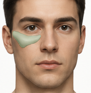
 The success of a custom infraorbital-malar implant depends far more on how it is designed than on the implant material itself. The objective is not simply to make the lower eyelid fuller, but to recreate the normal three-dimensional bony architecture that supports the lid-cheek junction. The implant should become an invisible extension of the facial skeleton rather than a discrete implant (bump of material on the bone).
1. Treat the Entire Skeletal Deficiency
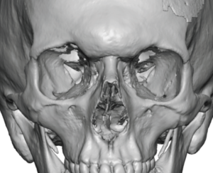
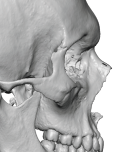
Undereye hollowing is rarely confined to the infraorbital rim. Most patients have varying degrees of deficiency which can be seen on a 3D CT scan involving:
- Infraorbital rim
- Medial malar eminence
- Anterior maxilla
- Upper zygomatic body
A successful implant should restore the entire deficient region instead of adding volume to only one isolated area.
2. Create a Continuous Lid-Cheek Transition
The most important aesthetic goal is eliminating the abrupt transition between the lower eyelid and the cheek.
This is accomplished by:
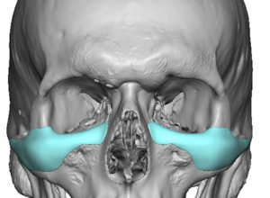
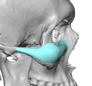
- elevating the infraorbital rim (saddling),
- extending onto the anterior malar surface,
- blending gradually into the surrounding bone.
The implant should create a smooth skeletal ramp rather than a raised ridge.
There are many variations to an infraorbital-malr implant design and each patient’s implant footprint will be different based on their aesthetic needs.
3. Differential Thickness
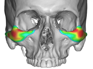
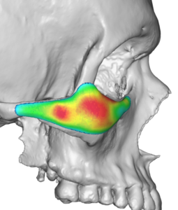
Unlike stock implants with a more uniform profile, custom implants vary in thickness throughout their surface.
Typical design characteristics include:
- greatest thickness directly beneath the tear trough,
- gradual taper superiorly toward the orbital rim,
- gradual taper inferiorly into the cheek,
- feathered lateral borders,
- smooth medial transition.
There should never be abrupt changes in implant thickness.
4. Respect Orbital Anatomy
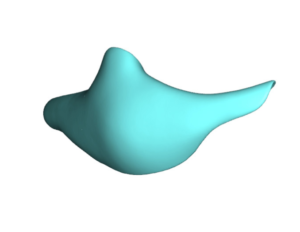
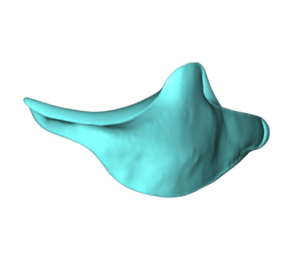
The implant should remain entirely outside the orbital cavity.
Important boundaries include:
- inferior orbital fissure,
- infraorbital canal,
- lacrimal drainage system,
- orbital septum.
The superior border should augment the orbital rim without projecting into the orbit or altering globe position.
5. Infraorbital Nerve Protection
The infraorbital nerve is the principal anatomic limitation.
Design considerations include:
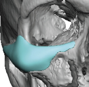
- identifying the exact foramen on the CT scan,
- incorporating a relief notch or channel when appropriate,
- minimizing the risk of compression over the nerve,
- avoiding screw placement near the foramen.
Failure to account for the nerve increases the risk of prolonged postoperative numbness.
6. Medial Extension
The medial portion of the implant deserves careful attention.
Excessive medial augmentation may produce:
- fullness adjacent to the nose,
- unnatural widening of the lower nasal bridge,
- excessive prominence near the medial canthus.
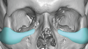
Most designs terminate just lateral to the lacrimal fossa while maintaining a smooth transition.
7. Lateral Extension
Laterally, the implant should continue beyond the orbital rim onto the zygoma.
This provides:
- improved cheek support,
- smoother contour,
- less visible implant edge,
- better integration with facial anatomy.
Stopping the implant too early often leaves a noticeable contour transition.
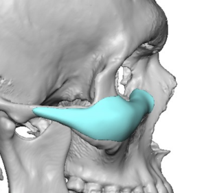
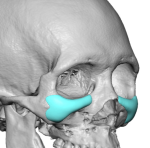
How far it shouild extend out onto the zygomatic arch will vary based on how much lateral cheek/arch augmentation the patient prefers.
8. Symmetry Does Not Mean Identical
Human facial skeletons are naturally asymmetric.
Custom implant design should correct meaningful asymmetries while respecting normal facial differences rather than creating perfectly mirrored implants. Independent right and left implant thickness maps often produce the most natural result.
9. Projection Direction
Volume alone is not the goal.
Projection should be directed:
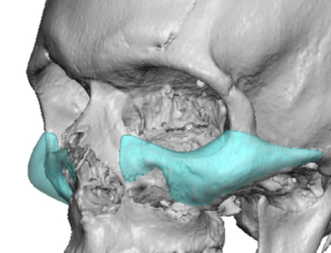
- anteriorly to support the lower eyelid,
- slightly superiorly to improve the lid-cheek junction,
- laterally to blend with the malar eminence.
Overemphasizing forward projection can create an artificial “cheek implant” appearance.
10. Negative Orbital Vector Correction
Patients with negative orbital vectors require more than simple rim augmentation.
The implant should:
- support the inferior orbital rim,
- advance the upper cheek,
- improve the orbital-to-cheek relationship,
- provide skeletal support beneath the globe.
This creates better lower eyelid support and softens the appearance of eye prominence.
11. Soft Tissue Considerations
The implant should complement, not overpower, the overlying soft tissues.
Factors influencing implant thickness include:
- skin thickness,
- subcutaneous fat volume,
- orbicularis muscle thickness,
- patient age,
- previous eyelid surgery.
Patients with thin soft tissues generally require more gradual contour transitions than those with thicker tissues.
12. Surgical Placement Requirements
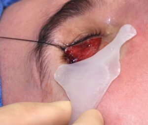
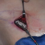
The design should facilitate straightforward surgical insertion. The best surgical access is through the lower eyelid NOT intraoral. Visualizing the infra-lateral orbital rim is critical for good placement and seeing it through the lower eyelid assures a higher likelihood of accurate implant placement.
Important features include:
- a single-piece implant whenever feasible,
- stable bony seating without rocking,
- predictable orientation,
- sufficient surface area for rigid screw fixation,
- borders that are easily identifiable during placement.
13. Avoid Overcorrection
Perhaps the most common design mistake is excessive augmentation.
The infraorbital region is one of the most visually sensitive areas of the face. Even small increases in projection can produce dramatic aesthetic changes. Conservative augmentation with smooth transitions almost always appears more natural than maximal volume enhancement.
14. Integration with Adjacent Facial Implants
When combined with custom facial implants elsewhere, the infraorbital-malar implant should be designed as part of the entire facial skeleton.
Its contours should blend seamlessly with:
- maxillary implants,
- paranasal implants,
- zygomatic implants,
- temporal implants,
- complete midface mask implants.
A comprehensive design strategy avoids visible transitions between adjacent implants and produces a harmonious enhancement of the midface.
Key Design Principle
The best custom infraorbital-malar implants are not recognized as implants at all. They recreate the patient’s missing skeletal anatomy by restoring a smooth, continuous transition from the lower eyelid to the cheek while respecting orbital anatomy, protecting the infraorbital nerve, and blending imperceptibly into the surrounding facial skeleton. The goal is not to make the cheek larger, but to restore the structural support that eliminates bone-based undereye hollowing and creates a natural, youthful lid-cheek relationship.
Dr Barry Eppley
Plastic Surgeon