
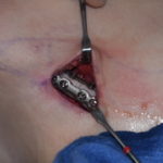
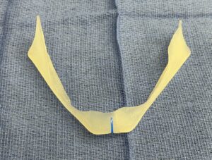
 For a custom jawline implant, the best results usually come from treating the problem as an inverse-design problem: define the desired postoperative external jaw contour first, then work backward to determine the implant geometry needed to produce it. The key is not simply designing an implant that “looks good” on the mandible. As some implant designs look great on the patient’s mandibular model but may not as significant external effect as one would have thought. Less commonly, but also a situation I have seen, is that the implant design may not look that profound on the implant model, but on the patient has a much more significant effect than the model design may indicate.
My approach is as follows:
Start with the augmentation goal in facial space.
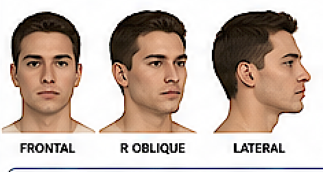
Use standardized photographs with with Photoshop or Ai imaging. Establish exactly where the patient wants change: chin projection/width, prejowl contour, mandibular body width, gonial width, angle definition, vertical ramus/angle length, or some combination. Frontal, oblique, profile, and submental views matter because an implant that achieves the desired profile can easily become too wide or unnatural from the front.
Create a target postoperative surface rather than drawing the implant directly. Ideally, digitally sculpt the desired external facial contour. Comparing this target surface with the patient’s existing surface produces a spatial “augmentation map”—essentially showing where additional contour is required and by approximately how much. This is substantially more informative than saying, for example, “add 6 mm at the angle.”
Translate soft-tissue change into skeletal/implant change. This is the difficult part. A 1-mm increase in implant thickness does not necessarily produce a 1-mm external contour change, and the relationship varies by location. Soft-tissue thickness, masseter position, periosteal elevation, implant edge geometry, skin/subcutaneous tissue and implant placement all affect the result. Therefore, a soft-tissue simulation or validated deformation model is preferable to simply subtracting the preoperative skin surface from the desired skin surface.
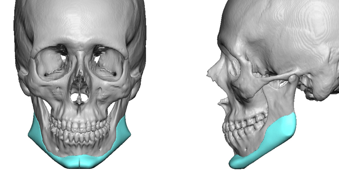
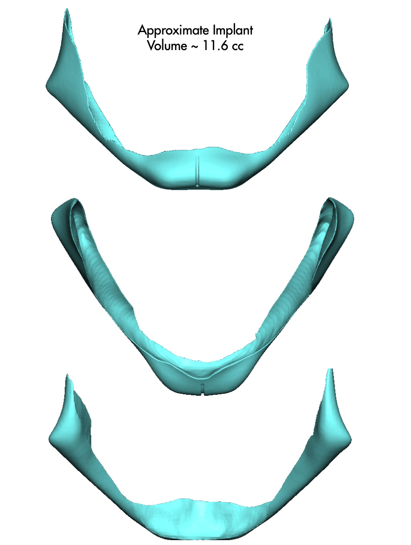
Design the implant against the actual mandibular anatomy. Once the required augmentation field is understood, create a patient-specific implant whose internal surface precisely conforms to the mandible while its external surface generates the required augmentation.
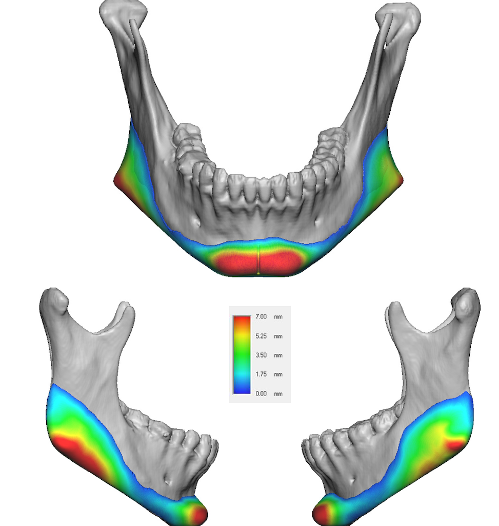
Evaluate thickness as a continuous 3D heat map rather than relying on a few linear measurements. Smooth transitions are particularly important anteriorly around the prejowl/chin interface and superiorly around the ramus/angle.
Iteratively simulate the result. The most useful design loop is implant – predicted soft-tissue result – compare with target – modify implant – repeat. Quantitatively compare the predicted and target surfaces with color-distance maps and cross-sections at reproducible anatomical planes. This makes the process much less subjective…although some subjectivity will exist.
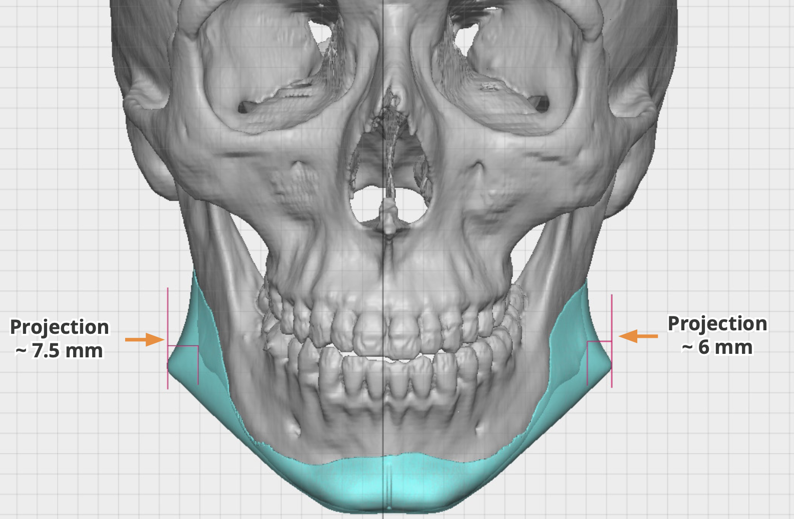
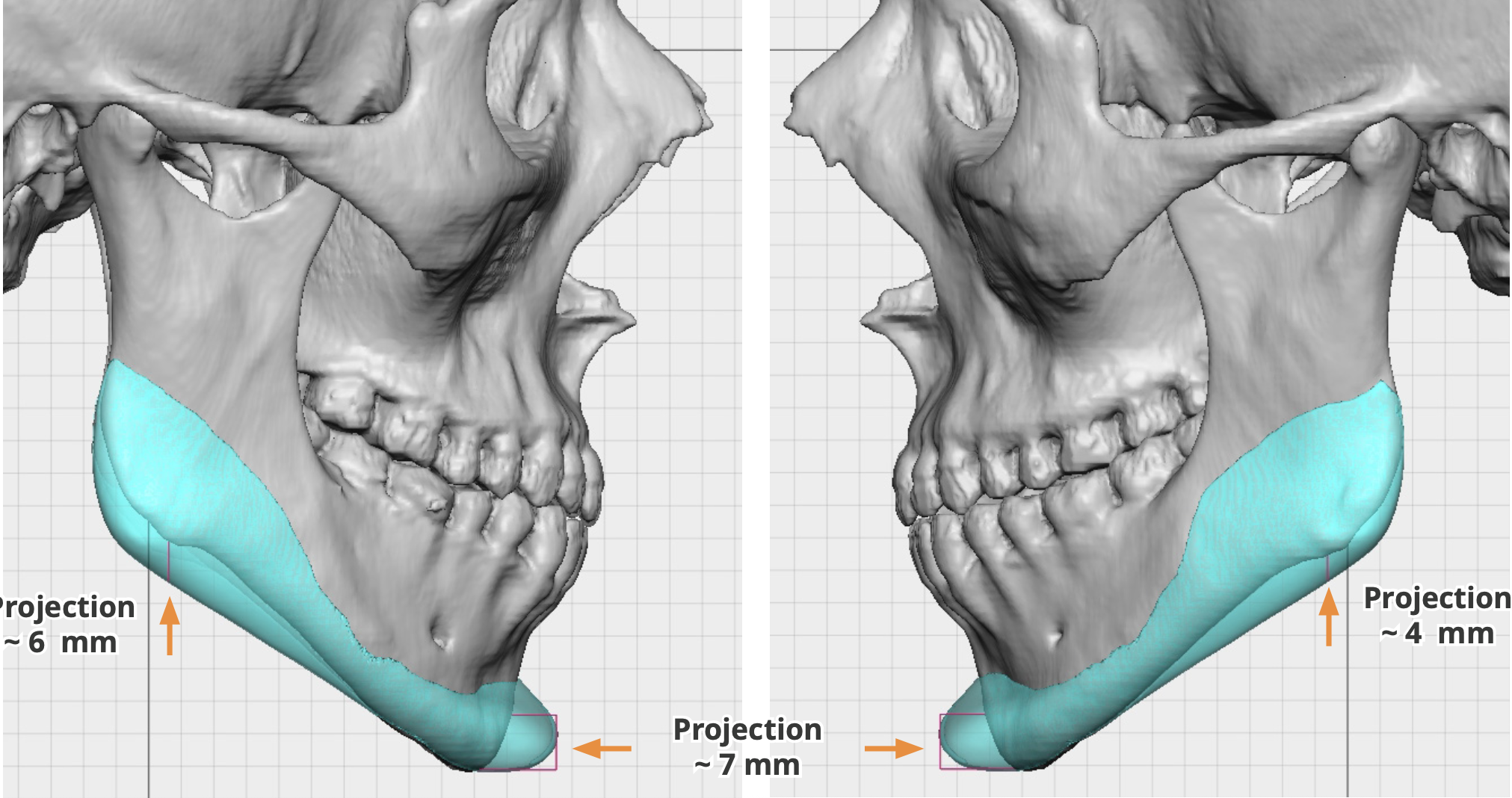 For total jawline implants, I would also separate the design problem into several controllable dimensions rather than treating “jaw augmentation” as one variable: chin projection, chin width/shape, anterior mandibular width, body width, posterior angle width, angle prominence, and vertical angle position/length. Those parameters interact, but separating them makes communication with the patient considerably more precise and the likelihood of achieving a pleasing result to the patient much more likely.
One particularly powerful approach would be a patient-goal-driven morphing workflow: generate several realistic 3D postoperative facial options first, let the surgeon/patient converge on the preferred contour, lock that surface as the design target, and then computationally solve for the implant required to approximate it. That reverses the conventional workflow of designing an implant first and then asking what the face might look like.
The major limitation is that the accuracy of the whole system depends on the soft-tissue response model. Bone-to-implant CAD is relatively deterministic; implant-to-final-facial-contour prediction is not. Consequently, I would put more effort into developing/validating the implant-to-soft-tissue transformation than into increasingly sophisticated manual implant sculpting.
Dr. Barry Eppley
Plastic Surgeon