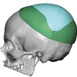
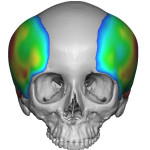
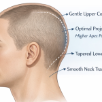
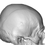
EXPLORE
Plastic Surgery
Dr. Barry Eppley

Explore the worlds of cosmetic
and plastic surgery with Indianapolis
Double Board-Certified Plastic
Surgeon Dr. Barry Eppley

Archive for the ‘skull reshaping’ Category

PMMA as an Aesthetic Cranioplasty Material

Sunday, June 9th, 2013


The use of skull reshaping with cranioplasty techniques that use synthetic materials dates back for over five decades. While the use of bone grafts is never an option in aesthetic skull procedures, it often is not an option in skull reconstruction either due to the size of the skull defect. This makes the use of alloplastic materials, often called bone substitutes or bone cements, as inevitable for many skull restoration procedures.

One of the historic and still most commonly used alloplastic bone substitutes in the skull is poly methylmethacrylate. (PMMA) Even though it is a polymer-based material and will never permit bone ingrowth or bone replacement, its low cost for its large volumes have always made it popular. Its widespread spread in joint replacements in orthopedic surgery as a true bone cement also speaks to its excellent biocompatibility.

Skull reshaping using PMMA has long shown it to be a well tolerated implanted material.   A recent published paper in the June 2013 issue of Aesthetic Plastic Surgery entitled ‘Aesthetic Refinement of Secondary Cranioplasty Using Methyl Methacrylate Bone Cements’ is interesting as it provides a more recent patient experience. Over a three year period the authors report on 20 patients who had PMMA implanted in their skull. Their use was reconstructive in nature for trauma and craniotomy patients and was used as a total inlay in the majority of the patients. (85%) The size of the cranioplasties was from 30 to 144 cm2 in size and involved implanted material volumes of 20 to 70 grams.

After an average two year followup period, no patients showed any evidence of implant infection, exposure, or extrusion. In addition, the PMMA reconstructions were structurally stable over this time period all the patients. The key to such successful outcomes in their observation were adequate and healthy overlying scalp tissues.

What does this mean to the aesthetic skull reshaping patients who has or is considering having PMMA implanted? This study is a small patient series that does not have significant long-term follow-up and used the material in an inlay rather than an onlay fashion. On the surface it would seem that this patient series has little correlation to aesthetic skull augmentations. But it does in one important way other than using PMMA…the scalp. All patients had overlying scalps that were healthy, not irradiated or missing tissue. Thus when re-expanded outward at the time of the reconstruction they developed no wound breakdowns or exposure.

PMMA is an effective and safe cranioplasty material. I have implanted far more PMMA in aesthetic patients than is in this paper for reconstructive purposes. I have been impressed with how well PMMA performs even though it is not remotely similar to its distant cousin, the hydroxyapatite bone cements. It does particularly well in the aesthetic skull augmentation patient undoubtably because the scalps are healthy and have never had surgery. Successful skull augmentation outcomes are predicated on normal scalp tissues because they will be stretched as the skull is built up underneath it.

Dr. Barry Eppley

Indianapolis, Indiana

The Small Incision Occipital Cranioplasty For A Flat Back of the Head

Tuesday, February 26th, 2013


Of all the non-craniosynostotic developmental skull deformities that exist, by far the most common is occipital plagiocephaly. This refers to a flattening on one side of the back of the head. While the deformity may be greatest on the flattest part of the occiput, its effects go beyond the flat skull area. Usually there is some compensatory bulging on the opposite normal side of the occiput as well as on the temporal side of the flattened side anterior to it. The ear positions can also be seen to be asymmetric with the ear on the flattened side more anterior than the opposite ear and may have some slight protrusion to it as well.

While a flat back of the head in an adult poses only an aesthetic deformity, to some so afflicted it can be more than just a casual asymmetric concern. While an occipital plagiocephaly in a balding male or one who shaves his head appears obvious and their concern is visible, I have seen an equal number of women with full heads of hair that are considerably bothered by it as well and adjust their hairstyles to accomodate for the aesthetic skull deformity.

The correction of an occipital flattening is done by building up the back of the head with a cranioplasty material. In and of itself, it is a fairly straightforward procedure to accomplish skull symmetry as long as one has complete visual access. Using a traditional and full ear to ear incision with peelback of the scalp does allow this exposure but many patients do not desire a full transverse scalp scar. This would be particularly true in almost all men due to their hair density and scar exposure concerns. It is always paramount to avoid trading off one aesthetic scalp/skull problem for another.

Adapting an occipital cranioplasty procedure through a smaller incision has been necessitated over the years because of scar concerns. It is possible to do the procedure but it necessitates several technical adjustments or modifications to that of a full open cranioplasty. It does not require special instrumentation but an intimate working knowledge of the handling properties of the various cranioplasty materials.

First and most importantly only one cranioplasty material has the working properties to be inserted through a small incision and molded into shape externally by scalp manipulation. PMMA or polymethylmethacrylate, acrylic bone cement, can be mixed into a putty which at a certain point in its set is not too loose but has not started to fully polymerize either. It is this window in the setting of the material that can permit it to be inserted through a smaller incision and still have adequate flow properties to be molded once inside. Unfortunately, none of the HA or hydroxyapatites have these working propertiues to be of great value for this approach. I have tried every HA material available and they all come up wanting, either in too short of set times or lack of adequate flow characteristics.

While a cranioplasty material can not really migrate around or away from its pocket on the bone, like other implants in soft tissues, some anchorage to the bone is always a good idea…even if it is just for psychological reassurance The best way to do that is to place small 1.5mm self-tapping titanium screws into the bone leaving them slightly above the bone surface. This will allow the PMMA to flow around them and lock onto them while it is curing. Since PMMA never really bonds to the skull bone, although there is some justification to calling it a bone cement since it does have some stick to it, this small screw fixation certainly prevents any micromovement. As long as too many screws are not placed or the screws are not too big, it is really quite easy to pop the implant off the bone later should that ever be necessary. In essence, their use does not make secondary removal unduly difficult.

Once the PMMA material is inserted, the scalp incision needs to temporarily stapled together. This then allows one to shape the material and feather its edges by external scalp manipulation. There usually is a few minute window to do the molding. While in years past the final set of PMMA was associated with very high heat release, this is no longer true. The exothermic reaction is very minimal with newer formulations so there is no risk of thermal injury to the scalp tissues. Once shaped and set, the staples are removed and partial visual assessment can then be done internally. (although this will be very limited as the material now occupies the entire pocket and the small incision makes it very hard to look over all the augmented area)

One advantage to the small incision cranioplasty is that the risk of difficulty with incisional closure is less because the molding is usually done away from it. This prevents the risk of encountering an incision that can not be closed due to the augmentation volume. When possible it is always best to have an incision in which no cranioplasty material lies underneath it. (ideal but not always possible)

The small incision occipital cranioplasty can be a very effective method of skull augmentation. Patients can have a very quick recovery and very minimal discomfort. But it is very technique dependent and requires good experience with more open methods of cranioplasty before attempting it through limited access.

Dr. Barry Eppley

Indianapolis, Indiana

Patient Testimonials: Skull Reshaping

Sunday, February 3rd, 2013

Dr. Eppley,

I just had to email you and the whole staff and thank you all for doing such a great job during my visit. This was the first time I ever had any type of surgery and we all know how nervous the word surgery can make a person. I was nervous the night before the procedure up until I walked into the surgery center. After being prepped by the nursing staff I was fine and there was no more worries. I’m still amazed at how it was done with little after affects. Dr. Eppley did a tremendous job and I thank him for that. The nurses that took care of me the day/night after the surgery were also great. I’m truly impressed with all that happened before, during and after the surgery. Please give my thanks to all those involved and I really appreciate all that was done for me.

Michael H.

Boston Massachusetts

Comment:

Amy form of surgery is a nerve wracking experience for any patient, but particularly so for a first timer from a distant city who is having a purely elective procedure. The importance of having a staff that is both trained and naturally inclined to be sensitive to these issues and help a patient through the process is the key to a good experience for the patient. While this is no less true than for a patient that lives down the street, the patient from a distant city or country is even more nervous as the very simple things, like sleeping and eating, have to be done in a very unfamiliar environment. Having had many patients come to Indianapolis from all over the world, we have developed a process for international plastic surgery that helps ensure an uncomplicated experience.

Skull reshaping surgery is usually surprisingly easy to go through with very little discomfort afterward. It should not be confused with a neurosurgery procedure where the head is shaved and the skull bones are removed. This is not how it is done at all. It is done on the outside of the skull bones whether it is adding a material or burring down some bone thickness. The bone itself has little to no feeling, it is the overlying soft tissues (periosteum and skin) that has the sensation. The raising of the scalp tissues will usually induce numbness over the elevated skin flaps so even this does not cause much discomfort. Most patients report postoperative discomfort more like a dull headache and many don’t take much pain medicine beyond the first few days after surgery.  If the skull work does not involve the forehead, then there is very little visible swelling as it is back in the hair-bearing scalp. This is why many skull reshaping patients can comfortably return home in just 48 hours after surgery.

Dr. Barry Eppley

Indianapolis, Indiana

Postoperative Instructions for Cranioplasty (Skull Reshaping)

Monday, January 28th, 2013


Skull reshaping surgery is done to create a more normal skull shape and contours. It achieves this goal through the application to the outside of the skull of either augmentation (by adding materials), reducing bone or a combination of both techniques. Most skull reshaping techniques require an open incisional approach to be done.

The following postoperative instructions for skull reshaping surgery are as follows:

1.  Skull reshaping surgery has a surprisingly minimal amount of postoperative discomfort. Pain medications are prescribed should you need them and you should take them as directed on the label, usually 2 tablets every 3 to 4 hour as needed. Many patients only use Tylenol; or Alleve after the first few days of surgery.

2.  There will be a circumferential head wrap applied at the end of the procedure. This is to be worn for the first night after surgery and can be removed the next day. Thereafter no dressings are needed. You may take it off the next day to shower.

3. In some cases of skull reshaping surgery, a drain will be used for the first day after surgery. This very small tube will be connected to a small bulb which collects any fluids. Empty the bulb as directed and there is NO need to measure the amount of fluid that comes out. In most cases of skull reshaping surgery, the bulb usually does not fill enough to be emptied more than once. The drain will be removed the day after surgery.

4. The scalp incision will be closed with either resorbable sutures, permanent sutures or small metal staples.There is no need to apply any antibiotic ointment to the incision, just leave them dry. Resorbable sutures do not need to be removed. Permanent sutures and staples will be removed 7 to 10 days after surgery. You may shower 48 hours after surgery and wash your hair.

5. You may wash your hair 48 hours after surgery. It is alright to get the sutures or staples wet. Dry and style your hair as desired. Be careful combing your hair so you do not catch the comb in the sutures or staples.

6. Strenuous physical activities and working out should wait for at least one week after surgery. While you can not harm the result by anything you do, wait until you feel better before exerting yourself.

7. You may eat and drink whatever you like right after surgery.  Focus on liquids and soft foods for the first few days after surgery.

8.   You may return to work and any non-strenuous physical activity as soon as you would like based on your comfort level.

9.   You may drive when you feel comfortable and can react normally and are off pain medication.

10. If any scalp or incisional redness, tenderness, or drainage develops after the first week of surgery, call Dr. Eppley and have your pharmacy number ready.

Consent for Plastic Surgery: Cranioplasty (Skull Reshaping)

Monday, January 28th, 2013


Every plastic surgery procedure has numerous issues that every patient who is undergoing a procedure should know. These explanations are always on a consent form that you should read in detail before surgery. This consent form, while many perceive as strictly a legal protection for the doctor, is actually more intended to improve the understanding of the various skull reshaping procedures. The following is what Dr. Eppley discusses with his patients for these procedures. This list includes many, but not all,of the different outcomes from surgery. It should generate both a better understanding of the procedure and should answer any remaining questions that one would have.

ALTERNATIVES

There are no alternatives to surgical skull reshaping. Some small skull defects may be treatable by a fat injections or bone cements placed through a minimal incision injection approach. High spots or skull reduction can not be reduced by a non-incisional approach.

GOALS

The goal of skull reshaping surgery is to create a more normal appearing skull contour. In some cases, this may require building up the bone, reducing raised areas or a combination of both reshaping techniques to get the optimal skull contour.

LIMITATIONS

The limitations of any cranioplasty procedure are how much of an incision can the patient tolerate (access and exposure), how much the skull can be built up based on the scalp’s ability to stretch and the thickness of the skull bone when reductions are being done.

EXPECTED OUTCOME

Expected outcomes include the following: temporary swelling of the scalp even extending down into the face, scalp skin numbness, a permanent scalp scar, the implantations of various biomaterials for augmentation/buildup including microcrew anchorage and months of healing and tissue settling until the final result is seen in all aspects. Healing of any cranioplasty procedure is a process and the minimal amount of time to judge the result is three months and may take as long as six months to see the very final result in many cases.

RISKS

Significant complications from skull reshaping surgery are very rare but could include infection. More likely but uncommon occurrences could include a wide scalp scar, potential hair loss along the incision, suture reactions along the incision edges causing local wound healing problems, edge demarcation/irregularities along any implant-bone interface, overcorrection of the skull contour, undercorrection of a skull contour, and asymmetries and irregularities of the skull contour. Any of these risks may require revisional surgery for improvement.

ADDITIONAL SURGERY     

Should additional surgery be required to revise a scalp scar, adjust a bone or implant contour or perform aesthetic adjustments of the initial skull reshaping will generate additional costs.

Case Study: Cranioplasty for the Triangular-Shaped Skull Deformity

Saturday, January 26th, 2013


Background: The appearance of the skull has a wide variety of shapes. But it most commonly has a front to back oblong shape that has curved round shape from the front that is deemed the most aesthetically pleasing. The shape from ear to ear across the top of the head should be a smooth convexity from the side muscular temporal regions transitioning across the bony top of the skull. This transition zone, known as the temporal lines, should not be too narrow to prevent loss of its convexity.

There are certain congenital skull conditions, known as craniosynotoses, that do affect the skull shape as it develops. One of these is sagittal craniosynostosis due to premature sagittal suture closure. This allows the skull to grow long in length but narrow in width. This results in a very narrow skull that often has a triangular or peaked appearance from the front view. Due to the severity of the skull defoemity, most of these affected individuals undergo early cranial vault reconstructive procedures to allow the rapidly expanding brain to help achieve a more normal skull shape.

But sagittal skull deformities do not always come in full expression and there are lesser degrees of their manifestations. Some would call these microform expressions of sagittal craniosynotosis. They can be very minor or more moderate in appearance. But they all appear with a a higher and and more narrow skull shape that creates a more triangular rather than a convex shape from the front view.

Case Study: This 26 year-old male had long been bothered by the shape of his skull. He had a very peaked skull shape that he felt was too high and too narrow. He did not like its very triangular shape which he likened to that of a house roof. He wanted a better skull shape so he could feel more normal and go one with the rest of his life without being embarrassed about his unusual looking head.

Under general anesthesia, a curved coronal incision was made across the top of the head from 1 cm. above each ear to the other side. Full-thickness scalp flaps were reflected in the front and the back to reveal the entire skull. The skull from above was very narrow and long with a raised sagittal ridge. Initially, a handpiece and burr was used to lower the sagittal ridge 5mm to 6mms from the forehead to the back of the head. Then the sides of skull were built up along the reduced sagittal ridge to the temporal line with PMMA material. Several 1.5mms titanium screws were placed one each side to anchor the PMMA material. All edges were then smoothed with a handpiece and burr. The scalp incision was then closed in two-layers using resorbable sutures.

He wore a head dressings and drains for the first 24 hours. Thereafter no dressings were used. he did eventually require needle aspiration of a seroma in the temporal area but this posed no long-term problems. He had substantial improvement in his skull shape, going from a triangular appearace to a more round convex form from the front view.

The peaked or triangular skull shape is often a manifestation of some form of sagittal craniosynostosis. It can be improved by a combination of lowering the ‘peak’ and filling out the side ‘valleys’. The limiting factor is that the narrow skull beyond the temporal lines can not be augmentted with material as it is covered with temporalis muscle.

Case Highlights:

1) The severely triangular-shaped head is usually the result of some degree of congenital sagittal craniosynostosis with a longer front to back length and a narrow skull width.

2) Skull reshaping of the triangular or peak-shaped deformity can be done by sagittal bone reduction, parasagittal augmentation or a combination of both.

3) A coronal incision is needed for wide open exposure to perform this form of skull reshaping.

Dr. Barry Eppley

Indianapolis, Indiana

Case Study: Surgical Reduction of the Occipital Knob Skull Deformity

Sunday, January 6th, 2013


Background: The shape of the skull frequently has various lumps and bumps of bone that stick up from an otherwise smooth surface. Many such bony protuberances have no specific reason for being in their location and are aesthetic nuisances that merely represent the way the skull formed and are present at a very early age. Other skull lumps are actually benign bony tumors, known as osteomas, that form for either no known reason or the result of trauma and calcification of a bone bleeding site. They occur later in life and are very discrete round elevations of bone which very commonly occur on the forehead and may be associated with very slow ongoing growth.

One very unique skull lump is the occipital knob. Occurring on the back of the skull at the junction of the bottom edge of the skull and the neck muscles, this is a skull bony protrusion that exists at the midline and can stick out just like a knob. Unlike developmental bumps or osteomas, the occipital knob occurs as an external occipital protuberance and is the result of a well known skull deformity. It usually occurs in patients who have a very narrow skull and reflects a mild manifestation of sagittal craniosynostosis. Premature closure of the posterior portion of the sagittal suture causes bone to heap up and develop an abnormal bony protrusion. The occipital bone becomes the site of compensatory overgrowth because it is located distal and perpendicular to the fused suture.

Sticking out like a knob, this skull protrusion can be very noticeable in men. Short hair cuts and shaved heads can reveal this abnormal skull protrusion. Like all skull lumps and osteomas, occipital knobs can be reduced to a normal skull contour. Because the occipital knob represents an abnormal thickening of skull bone, there is no risk of intracranial penetration or making the bone too thin in its reduction.

Case Study: This 45 year-old male had been bothered by a large protrusion on the back of his head since he was a child. It has been present as long as he could remember and had never changed in size. As he wore his hair shorter as he got older, it became more bothersome. He finally decided to have it reduced.

Under general anesthesia, he was placed into the prone position. Through a 6cm horizontal incision in the low occipital hairline, the very thick scalp tissue was elevated to expose it. A large bony bump was exposed that measured 3.5 x 3.5cm in diameters and was 2cms in height. A handpiece and burr was used to reduce the bony protrusion down to the level of the surrounding bone. The outer cortical bone was so thick that even in bringing it down to the surrounding skull contour the internal diploic space was not encountered and no bone bleeding was seen. The overlying incision was then closed after excising some excess scalp skin and underlying soft tissues. No drain was used.

A circumferential pressure dressing was used for the first 24 hours after surgery and then removed.  He took no pain medications and said he felt no discomfort whatsoever.When seen at one week after surgery, a small fluid collection (8ccs) was removed by needle aspiration to remove some slight fullness and return the scalp contour back to completely flat. He was thrilled to have solved his lifelong occipital knob concern so completely and easily.

Even at just one week after surgery, the occipital hairline scar was barely noticeable.

Case Highlights:

1)      An occipital knob is a midline protrusion of hypertrophic bone that sticks out at the base of the skull.

2)      Occipital knobs can be reduced through a small horizontal hairline incision to make for a smooth contour at the base of the occipital skull.

3)      For the bald, shaved or short hair styled men, a smooth skull contour is a desired aesthetic feature and can be achieved by a minor skull reshaping procedures for the occipital knob deformity.

Dr. Barry Eppley

Indianapolis, Indiana

Occipital Onlay Cranioplasty For Correction Of A Flat Back Of The Head

Friday, December 21st, 2012


Cosmetic skull deformities encompass a wide variety of concerns from flatness, pointy or high spots, ridges, bulges and asymmetries. But one of the most common head shape concerns is the flat back of the head. This specifically refers to varying degrees of lack of occipital projection, usually occupying an area between the top of the skull in the back down to the horizontal level of the upper portion of the ear. The causes are well known as a minor variant of occipital brachycephaly and often develop from early infantile positioning.

While flatness of the back of the head may seem trivial, to some affected it represents a significant aesthetic concern. The lack of a round posterior cranial shape is hidden by those concerned with caps and hairstyles and even devices that make the hair have more projection.  Some refuse to go swimming so their hair is not flattened to reveal an absent occipital roundness. Others feel their ‘flat head’ make them look unattractive and facially disproportionate.

The flat back of the head can undergo aesthetic improvement by a skull reshaping procedure known as an augmentation cranioplasty. Using either a plastic or hydroxyapatite material, an improved occipital shape can be obtained by building up the bony contour. Either material is applied in a putty-like state and manually shaped to the desired form until firmly set. Depending upon the incisional access and the amount of surgical exposure, the amount of cranial expansion can be up to 15mms to 20mms of augmentation if a competent scalp closure can be obtained.

A unique feature of most augmentation onlay cranioplasties, regardless of their location, is that the material must be applied to a smooth round surface. In addition, these round skull surfaces offer no inherent stability with most of their locations being the equivalent of on the ‘side of a cliff’.  As no known onlay cranioplasty material truly integrates into the underlying bone (or bone grows up into the implanted material), engagement of the material onto the skull’ surface has benefit. This is most conveniently done with small screw fixation.

Screw fixation of onlay cranioplasties, however, is not done as one envisions it for other facial implants. The implanted material is not first applied and then secured into position by screws. This is how it is done for preformed implants. Onlay cranioplasties are formed after they applied. Thus screws are initially placed and serve as a means of anchoring or something for the material to grab onto once applied, like metal rebar in concrete. This is particularly useful in occipital cranioplasties in which the material must be applied on the vertically-oriented back of the head where slippage and material displacement is very likely.

The pre-material placement of screws in onlay cranioplasty is useful for more than just implant anchorage. They are also helpful in setting the thickness of the applied material. By placing screw with lengths for the amount of thickness desired, the material can be applied using the screws as contouring guides.

Occipital cranioplasty can provide significant cosmetic improvement to those afflicted with a flat back of the head. Modern biomaterials such as titanium screws and acrylic and hydroxyapatite composites can very effectively create a more aesthetically pleasing occipital contour.

Dr. Barry Eppley

Indianapolis, Indiana

Case Study: Forehead Reconstruction with Hydroxyapatite Cement

Friday, September 28th, 2012


Background: Loss of the frontal or forehead bone can occur for a variety of reasons, usually from depressed fractures or loss of a craniotomy flap from infection. With removal of the protective bone cover, the brain and its dural covering sit directly up against the skin not only creating an obvious depression but pulsating with each heartbeat. Forehead reconstruction carries the highest aesthetic demands of any skull defect because it is the most visible in a non-hair bearing area and may involve the brow bone and brow ridge area.

There are almost a dozen methods of forehead skull reconstruction from split-thickness cranial bone grafts to computer-generated custom implant pieces. When skillfully done, any of these reconstructive methods will work satisfactorily. Their various advantages and disadvantages change based on the size of the forehead defect. The larger the bone defect becomes the more a synthetic approach becomes an appealing option.

One well established synthetic cranioplasty material for reconstructive use is hydroxyapatite. Consisting of the inorganic mineral content of natural bone, it is highly biocompatible although it does not get replaced by bone. It ends up creating a dense firm bone-like material that blends smoothly into the surrounding bone edges. It does not have the same strength as the normal double cortical layer skull bone but is strong enough to be an adequate skull substitute.

Besides the aesthetics of forehead skull defects, it is the only skull area which is contiguous with the air-filled frontal sinus cavity. This is a potential source of contamination and is a frequent source of forehead infections if a tissue layer is not created between it and the bone reconstruction material.

Case Study: This 13 year-old teen age boy was involved in a motor vehicle accident and sustained a severely depressed frontal forehead fracture and a large laceration down the center of his forehead. This required an urgent neurosurgical procedure with bone removal and repair of the dura. After three months of healing, he was left with a large depressed central forehead area (10 cm x 6 cm) that extended from the scalp down to the brows with a well healed vertical forehead scar. A 3-D CT scan shows the size of the defect and its involvement with the brow area and the frontal sinus.

Under general anesthesia, the forehead bony defect was accessed through his existing vertical scar from the scalp down to the area between the brows. The skin was lifting off of the dura and the surrounding bone edges. Near the brow area, the frontal sinus cavity was encountered as a 2cm x 2cm hole above the level of the dura.

The frontal sinus was clean and healthy with normal mucosal lining. A large pericranial tissue patch was sutured around all edges to create a thick tissue partition between the frontal sinus and the reconstruction site.

After the pericranial patch was placed, a floor was created for the reconstruction using titanium mesh. Thin 1mm titanium mesh was cut just larger than the bone defect and its edges were slipped under the defect to become a self-locking floor. This not only provided a containment method for the hydroxyapatite cement but keep the dural pulsations off of the hardening reconstruction.

Using a well known hydroxyapatite cement (Mimx, Biomet Microfixation, Jacksonville, FL), the activating liquid and calcium hydroxyapatite powder were mixed together into a putty consistency. This was then poured into the bone defect and molded into shape, recreating the lost brow bone area and the forehead above it.  The forehead skin was then closed and scalp scar removed prior to its closure in the hair area.

His surgery was done as an outpatient and he went home the same day. His head dressing was removed the next day and his sutures in the scalp removed ten days later. He had a smooth forehead result right with elimination of the forehead depression and the dural pulsations.

Case Highlights:

1) Reconstruction of the bony forehead can be done by a variety of techniques and hydroxyapatite is a well established cranioplasty material for full-thickness skull defects.

2) Forehead reconstruction which extends down into the brow area must take into account the frontal sinus and have a plan to keep it separate from any implanted material.

3) The properties of hydroxyapatite in a full-thickness skull defect needs reinforcement or a floor to add both strength and a containment method for the material.

Dr. Barry Eppley

Indianapolis, Indiana

Case Study: Cranioplasty for the Flat Back of the Head

Monday, July 23rd, 2012

Background:  The shape of the head is largely determined by the skull that lies underneath it.  Its normal oblong shape is created by the multiples plates of skull bone formed in utero that only formally fuse together after birth. The rapidly developing and expanding brain has great influence on forming the skull shape, much like an internal tissue expander.

But if the bone is blocked from being expanded, it will result in the push of the brain being directed elsewhere. This blocking effect can be created by a suture fusion (craniosynostosis) or an external force. By far, the most common cause of skull asymmetry is external molding. The most frequent form of external molding is inadvertent pressure caused by laying on one side of the head too long as a baby. This causes a classic flattening on one side of the back of the head with compensatory overgrowth on the other side. This is known as occipital deformational plagiocephaly.  On careful examination, one can often see from above that the entire head is twisted or rotated causing a cranioscoliosis effect in more severe cases.

With today’s shorter hairstyles and shaved heads, bothersome flat spots on the back of heads are becoming more evident. Often the ear on the flat side is moved further forward than the opposite ear and even the neck muscles on the flat side are asymmetric to the other side. These posterior skull asymmetries have given rise to patient’s requesting if they can be improved by some form of plastic surgery.

Case Study: This 42 year-old man was extremely bothered by the flat spot on the left side of back of his head. He had shaved his head for years since he began balding. To hide his concern about his flat spot, he always wore a hat to camouflage it. In discussing the option of a craniplasty correction, the issue of the trade-off of a fine scar was discussed. His level of concern about the back of his head made the scar issue a palatable exchange.

Under general anesthesia, a 9 cm horizontal posterior scalp incision was made. Wide exposure was made of both the normal and flat side of the occiput. Two small 1.5mm screws were placed at two different levels of the flat side marking how high the augmentation had to be for symmetry. Then using a PMMA acrylic cranioplasty material of 30 grams impregnated with antibiotic powder, a putty was made and inserted through the incision onto the flat occiput. The material was shaped through a combined internal and external methods until the area was both augmented and smooth and all edges were feather-like. The material set in ten minutes and the incision was then closed. A circumferential head dressing was applied and no drain was used. The length of the surgery was 90 minutes.

He had only minor discomfort the first night surgery and none thereafter. His head dressing was removed the next day. He had some expected swelling but no pain. Dissolveable sutures were used so removal was not necessary. He was placed on no restrictions after surgery and he could shave his scalp around his incision site 48 hours after surgery. His degree of occipital symmetry was dramatically improved.

Correcting a flat spot on the back of head is no different than a frontal or forehead augmentation. It is an onlay cranioplasty that requires a biocompatible material and an incision to place it. Scars are made as small as possible and material options are either an acrylic PMMA or HA. (hydroxyapatite) Cost plays a role in material choice. How much skull symmetry can be achieved is largely based on the volume added. The limiting factor in how much volume can be placed is scalp tightness and getting a good incision closure, preferably not directly over the implanted material.

 Case Highlights:

1)      One of the most common skull deformities is flattening on the back of the head, also known as occipital deformational plagiocephaly.

2)      Building up the flat back of the head is done by an onlay cranioplasty procedure, using a variety of different materials.

3)      Using an open approach, an occipital cranioplasty procedure is both very effective and involves minimal recovery.

Dr. Barry Eppley

Indianapolis, Indiana


Dr. Barry EppleyDr. Barry Eppley

Dr. Barry Eppley is an extensively trained plastic and cosmetic surgeon with more than 20 years of surgical experience. He is both a licensed physician and dentist as well as double board-certified in both Plastic and Reconstructive Surgery and Oral and Maxillofacial Surgery. This training allows him to perform the most complex surgical procedures from cosmetic changes to the face and body to craniofacial surgery. Dr. Eppley has made extensive contributions to plastic surgery starting with the development of several advanced surgical techniques. He is a revered author, lecturer and educator in the field of plastic and cosmetic surgery.

Read More


Free Plastic Surgery Consultation

*required fields


Pricing

The cost of any type of elective plastic surgery plays a major role in the decision to undergo the procedure(s).

Get Your Quote Here


My Plastic Surgery Story

Military Discount

We offer discounts on plastic surgery to our United States Armed Forces.

Find Out Your Benefits


Categories